WHAT WE OFFER
Diagnostic Imaging (DI) uses non-invasive technology to produce images of a certain area of the body. DI at Peterborough Regional Health Centre (PRHC) is responsible for many different types of medical examinations for inpatients and outpatients. We work to help determine a timely diagnosis and treatment for patients. Imaging tests performed at PRHC include:
Interventional Radiology
Computed Tomography (CT)
Magnetic Resonance Imaging (MRI)
Nuclear Medicine/Bone Mineral Density (BMD)
Ultrasound
Mammography (Breast Assessment Centre)
General Radiography (x-ray)
Picture archiving and communication system (PACS)
We offer 24-hour emergency coverage in x-ray, CT, ultrasound, and interventional radiology.
WHO WE ARE
Diagnostic imaging is made up of a multidisciplinary team that includes:
Radiologists
Nurses
Medical Radiation Technologist
Sonographer
Physicians
SERVICES
Interventional Radiology is a sub-specialty of radiology that uses a combination of radiographic imaging equipment, along with a radiologist to carry out diagnostic or therapeutic procedures. Needle biopsies and the placement of small tubes (catheters) to drain abscesses, blocked kidneys or bile ducts, are examples of what is done in the Interventional Radiology area.
Most procedures that are done are on an outpatient basis and can be successfully completed through a small incision in the skin that measures less than a quarter inch in diameter. The average times to complete a procedure depends on the type of procedure being performed.
PRHC has been offering computed tomography (CT) scans to the community since 1986. Our department currently has two CT scanners available to deliver optimal imaging services to both inpatients and outpatients. PRHC offers 24-hour emergency service to CT scans and can perform numerous exams including, but not limited to: head, neck, chest, abdomen, pelvis, and extremities.
Please note that some exams require an intravenous injection of contrast media (oral and/or rectal). The need for contrast materials is determined by the CT Radiologist. If a contrast medium is required, the patient will be informed when they receive an appointment time.
PRHC’s magnetic resonance imaging (MRI) centre provides this diagnostic service for the region. An MRI is a painless diagnostic examination that uses a strong magnet and radio waves to take cross-sectional images of the body without using x-ray.
Unlike x-ray or CT, there is no need for the use of ionizing radiation and there are no known side effects to having an MRI performed. Special circumstances involving pregnancy should be discussed with the ordering physician prior to requesting an MRI procedure. MRI exams can last between 15 – 60 minutes depending on the procedure selected and can involve a contrast medium being used – such as intravenously, and in some rare circumstances rectally.
Due to the special circumstances of an MRI procedure, there are several screening processes in place to ensure the safe examination of a patient. Prior to any appointment time being given, the patient and/or ordering physician must complete a medical history to ensure no contraindications (such as a pacemaker) are detected. Failure to properly complete the screening process will lead to delays in booking times and/or cancellation of the request.
Please note that specialty exams such as cardiac and prostate imaging are not offered at PRHC, please contact the MRI department for further questions regarding the type of exams offered.
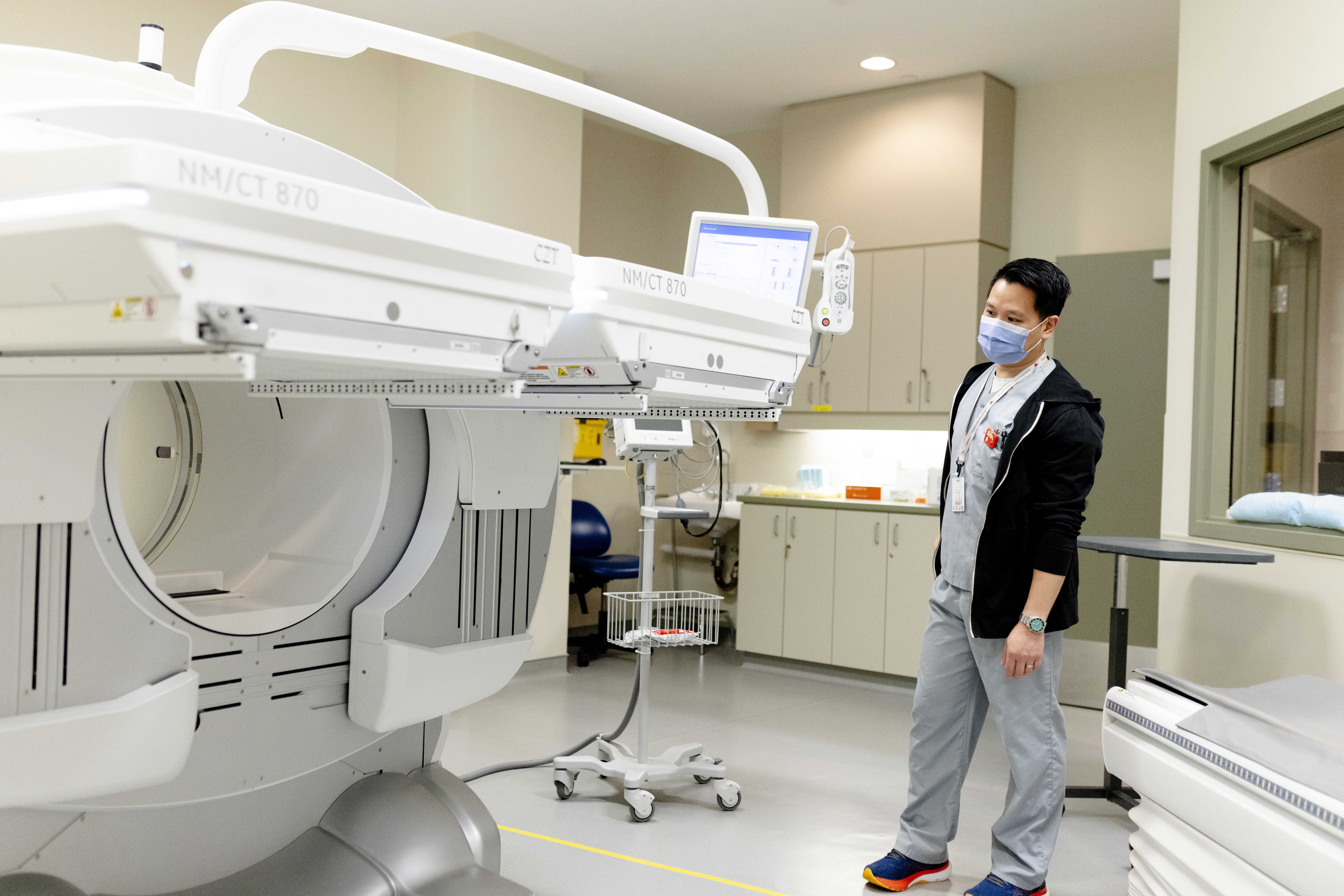
This division of Diagnostic Imaging performs diagnostic and therapeutic nuclear medicine procedures as well as bone mineral density testing. In diagnostic nuclear medicine, radiopharmaceutical is introduced into the body to target specific organs, bone or tissues. The radioactivity emits gamma rays from the patient’s body. These signals are mapped using highly specialized “gamma cameras,” creating an image on photographic film. Most nuclear medicine studies today are further processed with the aid of computer digitization and manipulation of these images.
Nuclear medicine scans provide unique information about both the structure and the function of major organ systems within the body.
The sensitivity of these studies means that the diagnosis can be made at an earlier stage in the progression of the disease. Therapeutic nuclear medicine is also performed in our department.
Bone Mineral Density (BMD) is a fast and simple procedure that evaluates and helps manage osteoporosis. BMD testing is performed using dual energy x-ray absorptiometry. A scan is taken of both the patient’s hip and lower spine using a small x-ray source. With input from the technologist, a computer analyzes the strength (density) of the bone from the images presented. Your doctor is then provided with a baseline measure of bone health. Osteoporosis patients may be treated, and the results of the therapy can be followed up with subsequent scanning.
The ultrasound department is an integral part of the Diagnostic Imaging department at the Health Centre. An ultrasound is an imaging method that uses high-frequency sound waves using a transducer to produce images of structures within your body. There is no use of ionizing radiation during an ultrasound procedure.
Imaging exams can be done on the following parts of the body: abdominal, renal, obstetrical, pelvic/trans-vaginal, pediatric, breast, scrotal, prostate (with biopsies), thyroid (with biopsies), and venous doppler assessments of veins in extremities.
Questions regarding accommodations (such as being in the ultrasound room during the procedure) should be directed to staff giving you the appointment.
HOW TO REFER TO DIAGNOSTIC IMAGING
Depending on the imaging test needed, you may need a referral from a physician or nurse practitioner or you may be able to self refer.


